CrystalsFirst

Categories
BLOG

EXPANDING THE TACABLE GENOME

EXPANDING THE TACABLE GENOME​

The DbTACs platform is a novel technology that enables selectively targeted protein degradation. It is designed to precisely degrade proteins of interest. The approach is compatible with various warheads such as small molecules, antibodies, or DNA motifs.

DbTCACs stands for DNA framework-based PROTACs. In comparison to PROTACs, the DbTACs platform uses a DNA framework-engineered chimera instead of a small molecule-based chimera, which provides a more stable and versatile platform for targeted protein degradation. Additionally, the DbTACs platform is able to target “undruggable” proteins and degrade proteins in a time-dependent manner, which are not features of PROTACs.

The platform uses a click chemistry-mediated programmable linker to achieve simultaneous selective multi-target proteolysis. The universality of the platform is highlighted by the following features: (1) precise degradation of POIs; (2) simultaneous selective multi-target proteolysis; (3) compatibility with various warheads; (4) a DNA framework-engineered chimera instead of a small molecule-based chimera; (5) a more stable and versatile platform for targeted protein degradation; (6) the ability to target “undruggable” proteins; (7) the ability to degrade proteins in a time-dependent manner; and (8) the ability to analyze the stability of the DbTACs using 2% agarose gel electrophoresis.

The DbTACs platform is a significant advancement in the field of drug discovery as it provides a new approach for selectively targeted protein degradation. The ability to precisely degradePOIs and achieve simultaneous selective multi-target proteolysis is a valuable tool for drug discovery. The platform is also compatible with various warheads, such as small molecules, antibodies, or DNA motifs, which provides versatility in targeting different kinds of proteins. The DbTACs platform can be used to develop new drugs for various diseases, including cancer, neurodegenerative diseases, and autoimmune disorders.

https://lnkd.in/eJZpFqxf

Categories
BLOG

UNCONVENTIONAL HALOGEN BOND DONORS

UNCONVENTIONAL HALOGEN BOND DONORS FOR DRUG DISCOVERY

CF2X moieties can be used as unconventional halogen bond donors drug discovery. They provide completely new opportunities to harness highly directional XB interactions. The CF2X structural motif is underrepresented in drug discovery and has hardly been applied so far, making it a promising area for exploration.

Vaas et al. propose that unconventional or unique binding modes could be explored based on CF2X-containing libraries, allowing for the discovery of unclaimed, patentable chemotypes and the establishment of added therapeutic opportunities. Their work also demonstrates that molecules containing C(sp3)F2X moieties attached by linker systems such as ethers or amides are synthetically accessible and that amide derivatives are particularly suitable for fragment-based drug discovery.

As an example, fragment 23 features an XB of CF2Br toward the P-loop, as well as chalcogen bonds, which are unique molecular interaction features. The implementation of CF2X acetamides into HEFLibs and biophysical evaluation (STD-NMR/ITC), followed by X-ray analysis, revealed these features and provided insights into the binding mode and interaction geometry of the fragment with the protein. This information can be used to design and optimize small molecules that target JNK3 and potentially other proteins with similar binding sites.

https://lnkd.in/e62hVTCr

PDB structure still to be released: https://lnkd.in/eES9pMJj
__
CF2X = X = Cl, Br, or I
JNK3 = c-Jun N-terminal kinase 3

Categories
BLOG

EXPANDING THE TOOLBOX OF COVALENT DRUG DISCOVERY

Expanding the toolbox of covalent drug discovery

Chiral sulfonyl fluoride probes can be used to directly map ligandable tyrosines and lysines in cells, providing a rich resource of liganded sites and the first reported clickable covalent probes for most of these sites.

Chen et al. provide a new approach for developing covalent chemical probes for potential therapeutic targets, which can be used in at least three ways:

(1) newly identified sites in potential therapeutic targets can become the focus of small-molecule screens

(2) sulfonyl fluorides can serve as chemical starting points for structure-based design of ligands with improved potency and selectivity toward selected sites

(3) sulfonyl fluorides can serve as clickable occupancy probes for cellular target engagement assays.

The probes also have an alkyne handle that allows for affinity enrichment and direct identification of covalently modified protein sites. A chiral 2-methylpiperazine amide linker provides stereoselective discrimination at the level of noncovalent and covalent binding.

Using these probes, Chen et al. identified hundreds of stereoselectively modified sites in functionally diverse protein sites, many of which lack existing chemical probes or drug leads.

Overall, this work provides a resource of ligandable tyrosines and lysines that can be used to develop covalent chemical probes. Among multiple validated sites, the researchers discovered a chiral probe that modifies Y228 in the MYC binding site of the epigenetic regulator WDR5. The chiral 2-methylpiperazine amide played a dominant role in molecular recognition, and the (R) enantiomer was consistently superior in labelling WDR5 Y228.

Paper: https://lnkd.in/e4PP9AMV

Crystal structure PDB 8F93: https://lnkd.in/eXksBZyy

Categories
BLOG

STRUCTURAL INSIGHTS AND COMPUTATIONAL MODELING DRIVE SELECTIVITY OF CRL4 CRBN RECRUITING PROTACS

Structural Insights And computational modeling drive selectivity of crl4 crbn recruiting Protacs

Small molecules inducing protein degradation are important pharmacological tools to interrogate complex biology and are rapidly translating into clinical agents. Selectivity remains a limiting challenge in the design of CRL4 CRBN recruiting PROTACs.

Bouguenina et al. used structural insights from known CRL4 CRBN neo-substrates, degron blocking design principles, and computational modeling to predict key interactions mediating the formation of productive ternary complexes.

The design principles on a previously published BRD9 PROTAC and generated an analogue with an improved selectivity profile. The computational modelling pipeline shows that the degron blocking design does not impact PROTAC induced ternary complex formation.

The computational modeling process was used to predict key interactions mediating the formation of productive ternary complexes. Molecular dynamics simulations were used to study the binding of the PROTACs to the target proteins and to predict the stability of the ternary complex. The application of free energy calculations estimated the binding affinity of the PROTACs to the target proteins. The design principles were applied on a previously published BRD9 PROTAC and generated an analogue with an improved selectivity profile, which was validated experimentally.

Categories
BLOG

PROTEIN DEGRADATION BEYOND UBIQUITIN PROTEASOME OR LYSOSOMAL SYSTEMS

Protein degradation beyond ubiquitin-proteasome or lysosomal systems

Targeted protein degradation (TPD) in eukaryotic cells relies on either ubiquitin-proteasome or lysosomal systems, which is powerless against target proteins in membrane organelles lacking proteasomes and lysosomes, such as mitochondria. The newly developed mitochondrial protease targeting chimera (MtPTAC) can specifically hydrolyze target proteins inside mitochondria.

Wang et al. have reported the development of a mitochondrial protease targeting chimera (MtPTAC) that can specifically hydrolyze target proteins inside mitochondria. The authors have validated the effectiveness of MtPTAC in inducing target protein degradation both in vivo and in vitro, using mitochondrial RNA polymerase (POLRMT) as a model protein.

The MtPTAC was designed as a bifunctional small molecule that can bind to mitochondrial caseinolytic protease P (ClpP) at one end and target protein at the other. Mechanistically, MtPTAC activates the hydrolase activity of ClpP while simultaneously bringing target proteins into proximity with ClpP.

For validation of POLRMT degradation, the researchers used quantitative PCR with reverse transcription (qRT-PCR) assay to detect the RNA level of POLRMT before and after treatment with MtPTAC. The assay indicated that MtPTAC did not decrease the RNA level of POLRMT at the concentration where the POLRMT protein level was significantly suppressed. This confirmed the effect of MtPTAC on protein reduction at the posttranslational level. The synthesized MtPTAC can effectively induce target protein degradation and the degradation efficiency is closely related to the PEG linker length.

This work demonstrates the powerful proteolytic ability and antitumor application prospects of MtPTAC, which could lead to the development of new therapies for diseases caused by mitochondrial dysfunction.

Paper: https://lnkd.in/e4ePQ6cD

Crystal structure of ClpP in complex with ONC201: https://lnkd.in/ezbSiGZn

Categories
BLOG

Review of the Drug Discovery Chemistry Conference 2023 in San Diego

Review of the Drug Discovery Chemistry Conference 2023, San Diego

by Dr. Serghei Glinca

The challenge of the industry remains – access to chemical matter. The DDC conference was full of highlights focusing  mainly on enabling technologies and platforms that unlock chemical matter.

Fragment-based drug discovery (FBDD)

The range of applications of FBDD ranged from non-covalent to covalent screening techniques. One of the best talks highlighting the application of crystallographic fragment screening on KEAP1 was by Marcel Verdonk. I want to emphasize that in this work the FBDD campaign was used in a versatile way. Crystallographic hits, although exhibiting low affinity, were used as starting points for fragment linking. The resulting compounds with lead-like properties were used to build up a focused HTS-library.

This results in a nM compound. Stephen Fesik highlighted how to address the transcription factors Myc by targeting WDR5, which resulted in potent compounds which have been validated in animal studies. It seems that the beta propeller proteins are an emerging target family that are well amenable for FBDD.

Covalent Drug Discovery

The covalent space has been growing significantly over the past years, which is reflected by 24 talks at the conference. Dan Nomura has highlighted the application of chemoproteomic platforms using covalent fragments. It’s striking that covalent labeling leading at IDPs induces an even higher disorder leading to protein degradation.

This has been demonstrated for Myc by targeting the intrinsically disordered Cys171. Prof. Pellecchia highlighted that going beyond cysteines is a viable strategy for gaining selectivity targeting e.g. lysines. Tuning experimental setups to enable covalent labeling of lysines seems to be critical for high-quality results. For example, sulfonyl chlorides can label lysines but can also react with the His-tag, which depends on the concentrations of the ligand vs. protein.

It seems that library screening without the His-tag is a better idea. Joachim Broeker from Boehringer Ingelheim presented how BI-0474 was developed. FBDD and SBDD were enablers. The interesting part was that they grew their compounds by starting from a covalent screen of fragments of the S39C mutant. The fragments were bound to the switch II pocket and used the hits as non-covalent hits and grew them towards the Cys12, leading towards the reversible covalent inhibitor BI-0474

DNA-encoded Libraries (DEL)

Although I have not been participating at the DNA-encoded libraries (DELs) track, it seems that DEL screening is gradually replacing the “gold standard” to screen for Ro5 compounds in HTS. In discussions with colleagues, I learned that some companies are taking advantage of DEL screening, which enables access to a larger chemical space compared to HTS. The field is still evolving and Prof. Joerg Scheuermann from ETH showed the potential of the DEL technology for macrocycles. We’ll see more developments from the DEL space.

AI for Drug Discovery

Due to the overlap with other talks I could only join several talks of the AI for drug discovery track. Most of the AI/ML applications are still for relatively basic scenarios but machine learning complements drug discovery technologies quite well. The question that I often ask is whether we are better off with ML-based tools or with “just” executing the experiments and synthesizing molecules. Of particular note was the presentation by Bryce Allen from Differencitated Therapeutics.

It was quite impressive how the preference for ligases of specific compounds can be predicted by their engine. Also, a target id case study by InSilico Medicines showed how fast hypotheses can be generated and tested using AI platforms, which was demonstrated for CDK20. I believe this is the strength of the AI/ML platforms, namely, generation of a higher number and potentially more precise hypotheses for experimentation. 

Overall, FBDD, AI and DELs are really exciting technologies that will deliver even more exciting drug discovery stories in future.

Categories
BLOG

The Protein Data Bank has over 200,000 structures.

The Protein Data Bank has over 200,000 structures.

Structural biology is an important field in drug discovery as it helps researchers understand the three-dimensional structure of proteins and how they interact with other molecules. This knowledge is crucial in developing new drugs as it allows scientists to target specific proteins and understand how they contribute to diseases. 

One of the most important resources in structural biology is the Protein Data Bank (PDB). The PDB is a database that contains the 3D structures of thousands of proteins, which can be used by researchers to study the structure and function of these proteins. The PDB is a valuable tool for drug discovery as it allows scientists to analyze the structure of disease-causing proteins and identify potential drug targets. 

1. The deposited structural data helps to develop new drugs and supports biomedical research

The PDB allows for the comparison of different protein structures, which can help researchers identify common structural features that are important for disease progression. By understanding these features, scientists can develop new drugs that target these specific regions, which can help to improve the effectiveness of treatments. 

The publication by Goodsell et al. 2010 reported that 90% of the 210 new therapeutics approved by the US Food and Drug Administration (FDA) between 2010 and 2016 were discovered partly due to the PDB archive holdings. 

2. Database for AI/ML training

The public access to the PDB data laid the groundwork for the development of Artificial Intelligence and Machine Learning methods for predicting protein structures. Currently, the collection requires more than 1 TB of storage and containing more than 3,000,000 files associated with these PDB entries.  

In addition to providing structural information, the PDB also contains information on protein-ligand interactions, which can be used to understand how drugs bind to their targets. This information can be used to design new drugs that specifically target disease-causing proteins, improving their efficacy and reducing side effects. 

3. Over 50 years of research

Since its inception in 1971, structural biologists from all over the globe have contributed their experimentally determined protein and nucleic acid structure data. It is because of their efforts that this central, public database has attained this important milestone. 

We say thank you to all contributors and structural biologists who put tremendous efforts in solving 3D structures and achieving the milestone of 200,000 structures 

Categories
BLOG

X-RAY CRYSTALLOGRAPHY: PROTEIN SAMPLE REQUIREMENTS

X-Ray Crystallography: Protein Sample Requirements

A precondition for a successful crystallographic screening is a pure, stable, and monodisperse protein sample.

In the process of protein production, controls such as SDS-gels already indicate towards the purity of a protein sample. Additionally, during standard concentration determination via UV/VIS, contamination by DNA or RNA can be assessed. In the following section, typical methods for assessment of protein quality are described with the focus on crystallography-grade protein quality. 

After protein production and before crystallization it is essential to check for protein quality. Initial assessment via native Mass-Spec can confirm the correct size of the purified protein with more precision than a SDS gel and can also give first indication regarding oligomerization states or very tightly bound cofactors as well as the presence of posttranslational modifications. Even if the purified protein has the correct molecular weight, it can still be unclear whether the protein is correctly folded.  

Protein folding is a key process in biology since it is ultimately responsible for their biological function. Proteins that are misfolded or not folded at all may lead to unsatisfactory results in subsequent experiments or significant loss of protein material due to aggregation over time. Ultraviolet circular dichroism (CD) is the method of choice to monitor changes of protein structure in solution providing information of  secondary protein structure, hence, the correct folding. 

Protein samples need not only to be correctly folded but also stable and monodisperse. Thermal shift assay (TSA) measures the melting temperature of a protein (Tm), which is an indication of protein stability. Several factors such as pH, salt, cofactor, or buffer composition influence protein stability. Ideally, the protein is measured and stored in a buffer where it is more stable.  

Melting curve of T. cruzi FPPS protein: the addition of Mg2+ increases protein stability by about 5°C (Francesca Magari)

Proteins also exhibit different stability against freeze-thaw cycles. This stability is a very important factor in inter-lab and general process optimization and can be improved via buffer optimization. Proteins that are not stable against freeze-thaw cycles have to be crystallized immediately after purification and cannot be sent on dry ice. The lifetime of such proteins can usually be extended via storage at 4°C or on wet ice but is much shorter than at -80°C. However, there are some rare cases in our experience, in which storage at lower temperatures is disfavored and the protein must be kept at room temperature or above. 

Protein can be stable in a certain buffer, but the composition of the sample is not necessarily monodisperse. Monodispersity means that the protein exists in solution as a single oligomeric species, i.e., monomer or dimer, and is free of non-specific oligomers and aggregates. This can be checked with dynamic light scattering (DLS). 

In summary, the quality of crystallography-grade protein material is significantly higher compared to assay-grade protein. A checklist for crystallography-grade protein samples are

  • free from DNA/RNA
  • stable: Tm > 30 °C
  • monodisperse
  • properly folded
  • resistant to freeze-thaw-cycles
  • high purity according to analytics
Categories
BLOG

FRAGMENT HIT IDENTIFICATION IN FBDD

Fragment hit Identification in FBDD

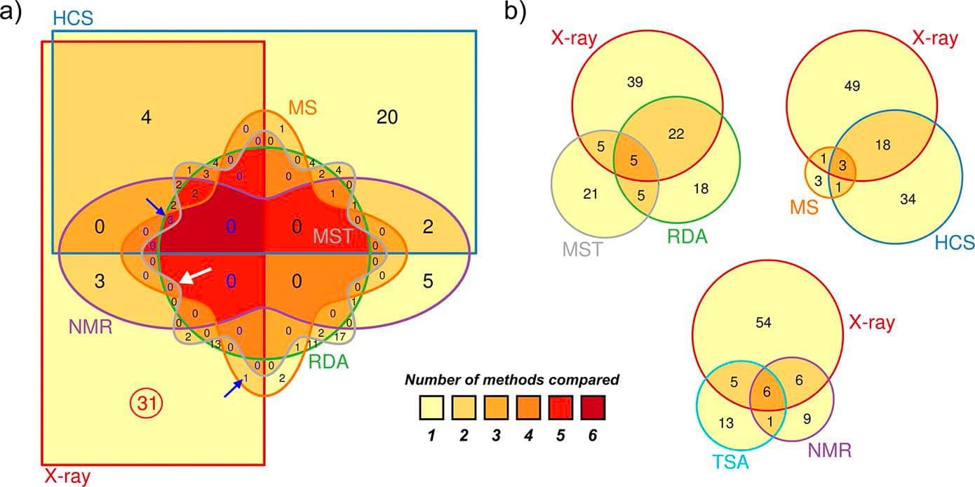
In the field of fragment-based drug discovery (FBDD), biophysical methods such as NMR, native MS, high-concentration biochemical screens (HCS), thermal shift assays (TSA), or surface plasmon resonance (SPR) are usually used as pre-screening techniques to screen fragment libraries to identify the most promising binders.  

However, employing a subsequent cascade of these methods prior to X-ray crystallography has been proven rather misleading as they might miss an important fraction of binding fragments that could be observed in crystal structures. In addition, the overlapping hits resulting from biophysical methods compared to X-ray crystallography have been rather poor.  

Venn diagrams show low overlaps between hits identified in X-ray crystallography and other biophysical methods.

Why not a direct crystallographic fragment screen?

Among all possible screening methods, X-ray crystallography is not only the most sensitive method for detection of fragment is binding. It also reveals the geometry of binding as precise three-dimensional positions of atoms in a protein structure. This is essential to investigate the accessible chemical space in a protein-ligand complex for further development of the initial hit into a lead candidate. Highly resolved crystal structures can also reveal the exact position of water molecules and water networks in the binding site. In this regard, it has been shown that water molecules in fixed position and their displacement can be an important data in structure-based lead discovery.  

Remarkably, X-ray protein crystallography is not only able to detect high- but also low-affinity binders that cannot be detected by any other method.  

Only after having precise structural information about the initial hit and exploring the binding pocket, other biophysical methods can be used to further characterize the protein-ligand complex and improve affinity, potency, and binding kinetics of fragment expansion campaigns.  

Osborne, J.; Jhoti, H. et al., 2020

High-quality crystallization and high-performance soaking systems

Thanks to third-generation synchrotron sources and the latest methodological improvements in automated crystal mounting systems, data collection, and processing, it has become increasingly feasible to screen entire libraries of fragments efficiently. Although automation enables efficient data collection, the most important part before data collection is the development of a high-quality crystallization and high-performance soaking systems.  This could be compared to assay development for high-throughput screening campaigns. The quality of crystals in terms of diffraction and reproducibility has to deliver consistent and comparable results. It’s not about getting one crystal at sufficient quality, but rather hundreds of crystals, which enables a screening at all. This requires specific expertise, technology and a broad range of experience.  

With novel workflows and pipelines, like FastForward, it is possible to utilize the large amount of collected data in a smart way. In fact, starting from the raw data given by X-ray protein crystallography, it is possible to process and refine the datasets of fragment library screens toward high-quality 3D models to design higheraffinity compounds in a short period of time.   

Subscribe to stay tuned